The Importance of Measuring Muscle Activation

By |2024-03-13T13:01:41-04:00March 31st, 2024|Latest Articles|

One of the biggest benefits of surface EMG biofeedback is its ability to measure the muscle activation of common exercises provided by rehab professionals every day. Thanks to an abundance of research on sEMG activation patterns during specific exercises, rehab providers can use an evidence-based approach for their exercise selection.(1,2,3) However, just because research has demonstrated the best exercises for the activation of the gluteus medius or gluteus maximus, doesn’t mean our patients are performing the exercise in a manner that elicits the desired EMG activation levels.

The body is extremely good at accomplishing a given task, no matter the cost. When the primary muscle responsible for a certain movement becomes too weak or fatigued, the synergistic muscle will step up to get the job done.(1) For example, when the glute med and glute max become weak, it is common for activation levels of the tensor fascia latter (TFL) to significantly increase.(1) While this is a valuable characteristic for our functional performance as humans, when this compensatory movement pattern persists for an extended period of time, working back into the correct movement pattern, with the correct primary muscle activation, is a challenge. Compensatory movement patterns are a very common side effect of pain or injury. One of the best applications of mTrigger biofeedback is that it allows rehab professionals to really focus in on how their patients are performing their exercises.

Let’s look at exercises the literature supports for the gluteus medius and gluteus maximus under desired and “healthy” conditions.

In three different studies, gluteus activation was tested from three different perspectives.

The first study (1) looked at the COMPARISON of glute medius, glute maximus, and TFL activation during several common hip exercises. Here are some notable findings:

  • Clamshells with and without a resistance band produced a higher amount of glute max activation, then glute med, then TFL.

  • Sideying hip abduction produced a high amount of glute med activation, then glute max, then TFL.

  • Standing hip abduction yielded high glute med and TFL activation followed by glute max on both the standing and moving legs.


The second study(2) investigated how the DIRECTION OF APPLIED FORCE effected muscle activation of the glute med and glute max. For instance, how does the EMG muscle activation levels of glute max differ between a squat or lunge (vertical force) and a deadlift (horizontal force). Some important findings included:

  • Glute max activation is much higher when a horizontal force is applied compared to a vertical force. In fact, when performing a 1RM squat or trap bar deadlift and a 1RM hip thrust, the activation of the glute max is 16% higher in the hip thrust exercise.

  • Vertical force exercises that create a very high glute max activation include skater squat and a single leg wall squat (with the other leg extended).

(Skater squat video)

  • Horizontal force exercises that create a very high glute max activation include a plank with bent knee hip extension, prone hip extension with upper body on table and flexed contralateral knee joint on a chair, single leg bridge with knee bent to 135 deg and foot flexed, and a single leg bridge with 90 deg of knee flexion and foot flat

In a third study(3), researchers sought to find the HIGHEST LEVELS OF EMG activation of the gluteus medius during exercises. Here is what researchers found:

  • Very high levels (60%+ MVIC) of muscle activation were seen during the following exercises: step up, lateral step up, diagonal step up, crossover step up, barbell hip thrust, hex bar deadlift, split squat, and a lunge.
  • Different types of squat variations (stance width, depth of squat, barbell position) all effected glute max activation levels.


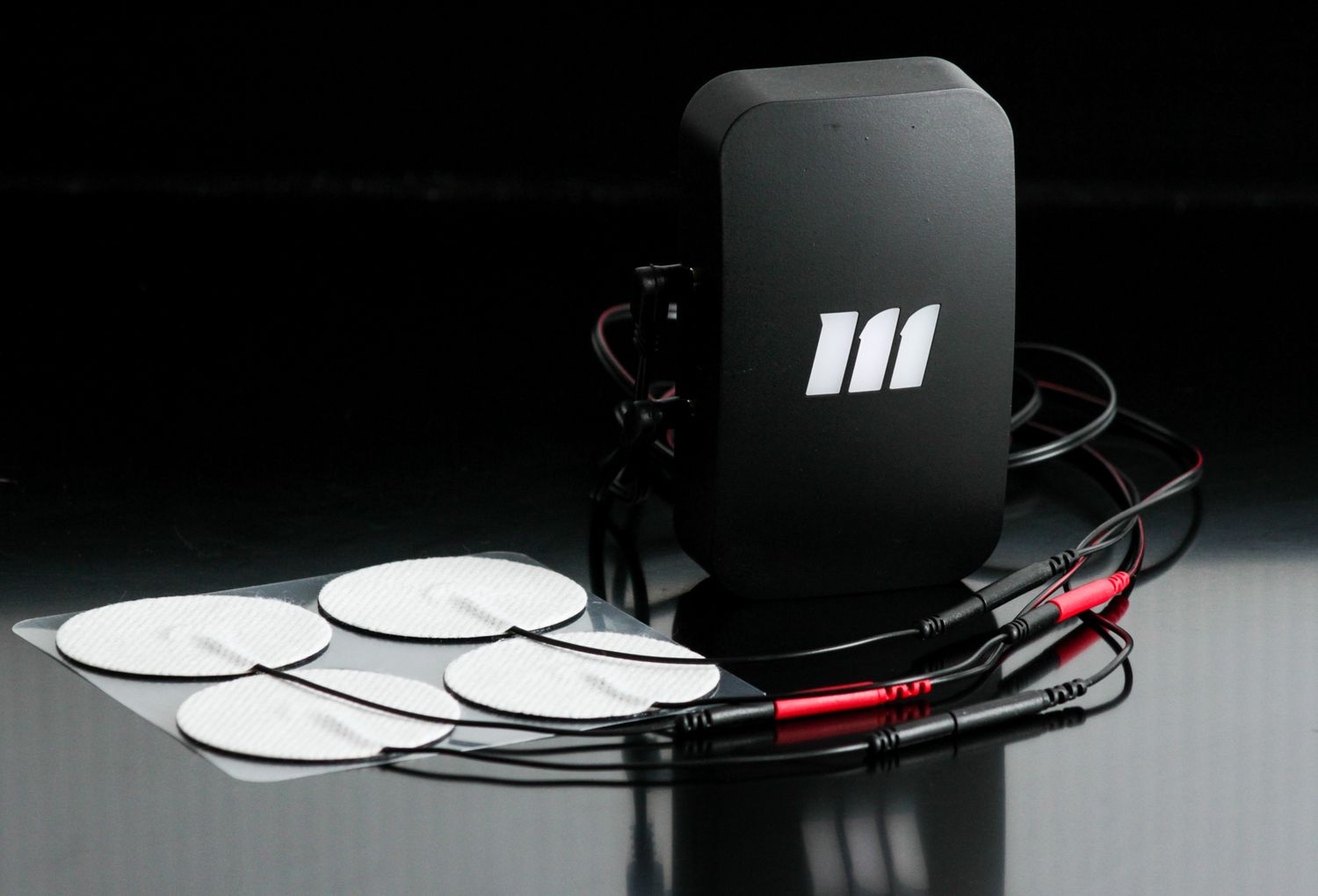
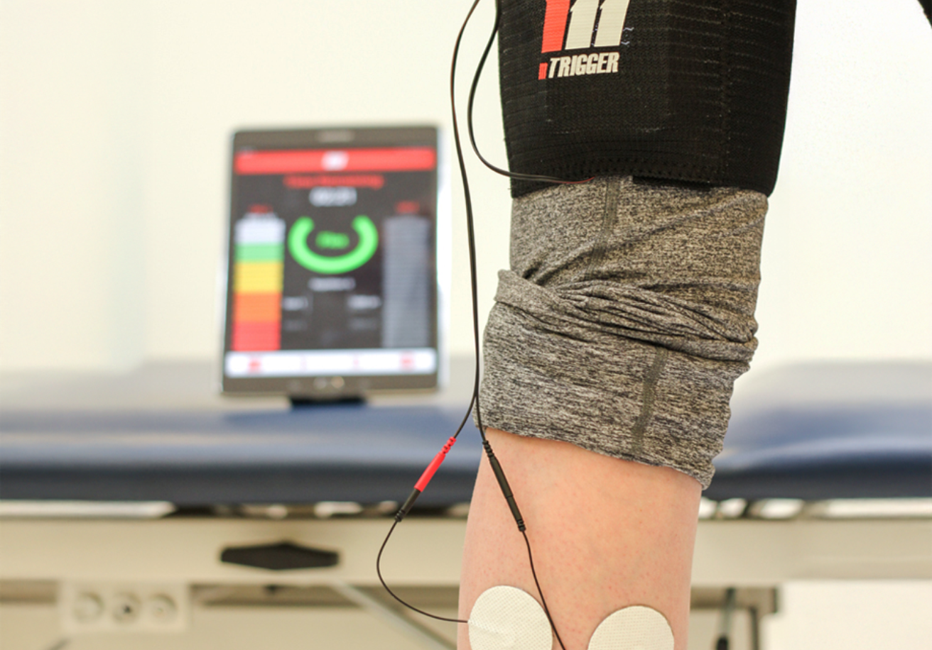
Equipped with this knowledge, you can now use the mTrigger biofeedback device to really see what muscle activation looks like in your patients compared to what you might expect. From the research by Neto et al. we expect to see a high level of glute max activation during a step up.(3) In this athlete, who is experiencing low back pain, the activation level of the glute max is very low compared to what we expect to see, despite the exercise “appearing” to be performed very well. We know from Macadam et al, that low back pain is commonly associated with weakness and strength imbalances in the glutes.(2) This is exactly what we are observing here, however, without the use of mTrigger biofeedback, you wouldn’t be able to ‘see’ the compensation patterns.

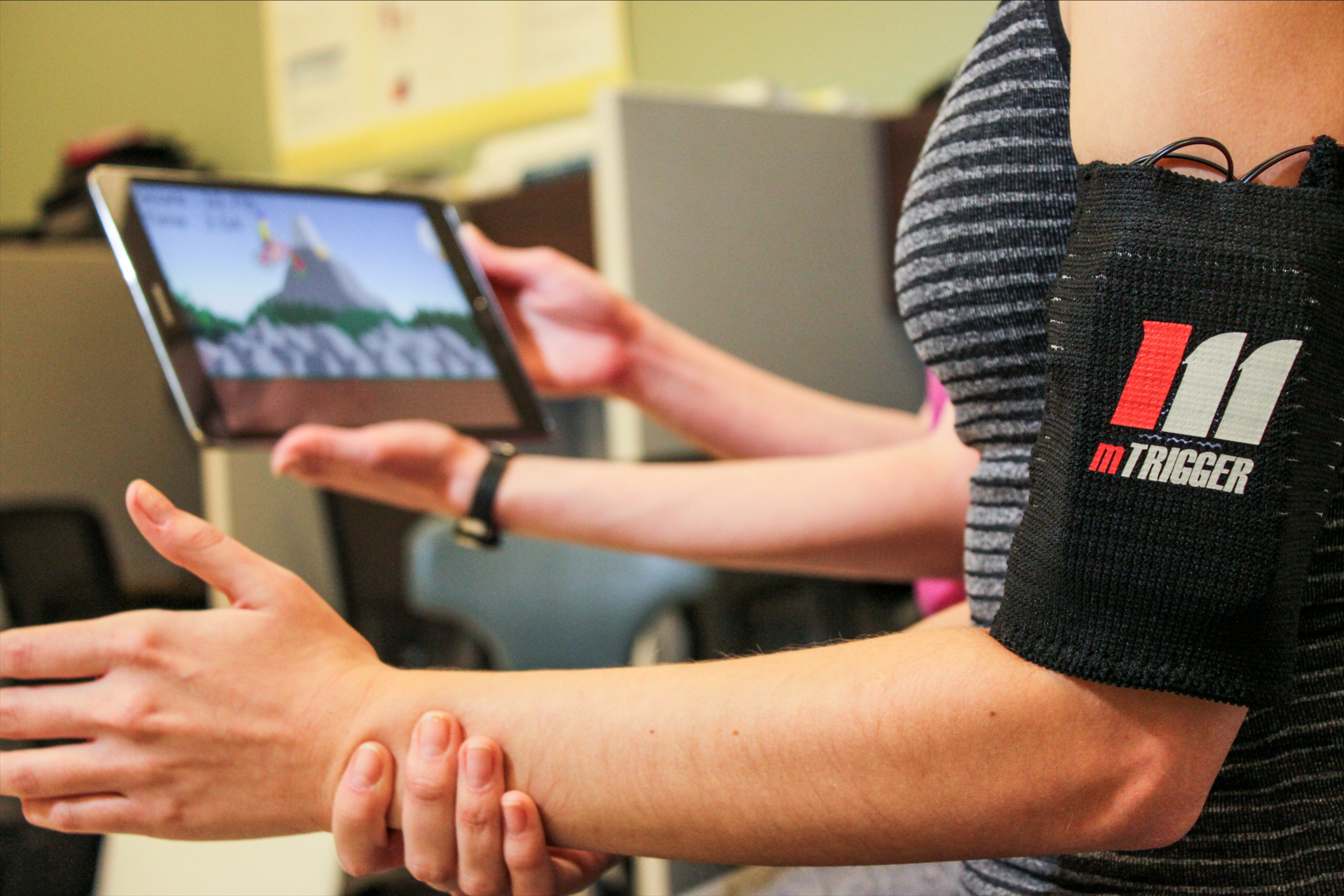
Once identified, the use of mTrigger biofeedback arguably becomes even more critical. Now visual biofeedback can be used to help the patient make changes to their movement patterns to elicit the desired and correct muscle activation.

Summary
Understanding what muscles are activated during commonly prescribed rehab exercises and to what extent, helps providers better select exercises to match the capabilities of the patient in front of them. Using mTrigger biofeedback helps to provide insight into how the neuromuscular system is functioning and should be used to help guide exercise selection and monitor patient performance. Selecting the best exercise or putting together the “best” program, has very little effect when the exercises are not done correctly. By combining data of EMG activation levels with the use of mTrigger biofeedback, you will begin to see the effects your evidence-based exercise selection has on your patient outcomes.


Reinforcing Proper Movement Patterns 

READ MORE HERE


Training Eccentrics with mTrigger Biofeedback

CLICK HERE


 References

  1. Bishop BN, Greenstein J, Etnoyer‐Slaski JL, Sterling H, Topp R. Electromyographic Analysis of Gluteus Maximus, Gluteus Medius, and Tensor Fascia Latae During Therapeutic Exercises With and Without Elastic Resistance. Int J Sports Phys Ther. 2018;13(4):668. doi:10.26603/ijspt20180668
  2. Macadam P, Feser EH. EXAMINATION OF GLUTEUS MAXIMUS ELECTROMYOGRAPHIC EXCITATION ASSOCIATED WITH DYNAMIC HIP EXTENSION DURING BODY WEIGHT EXERCISE: A SYSTEMATIC REVIEW. Int J Sports Phys Ther. 2019;14(1):14. doi:10.26603/ijspt20190014
  3. Krause Neto W, Soares EG, Vieira TL, et al. Gluteus Maximus Activation during Common Strength and Hypertrophy Exercises: A Systematic Review. J Sports Sci Med. 2020;19(1):195.  

Share This Article

Leave A Comment

Title

Go to Top